In the following you can see research methods and devices we use for our research
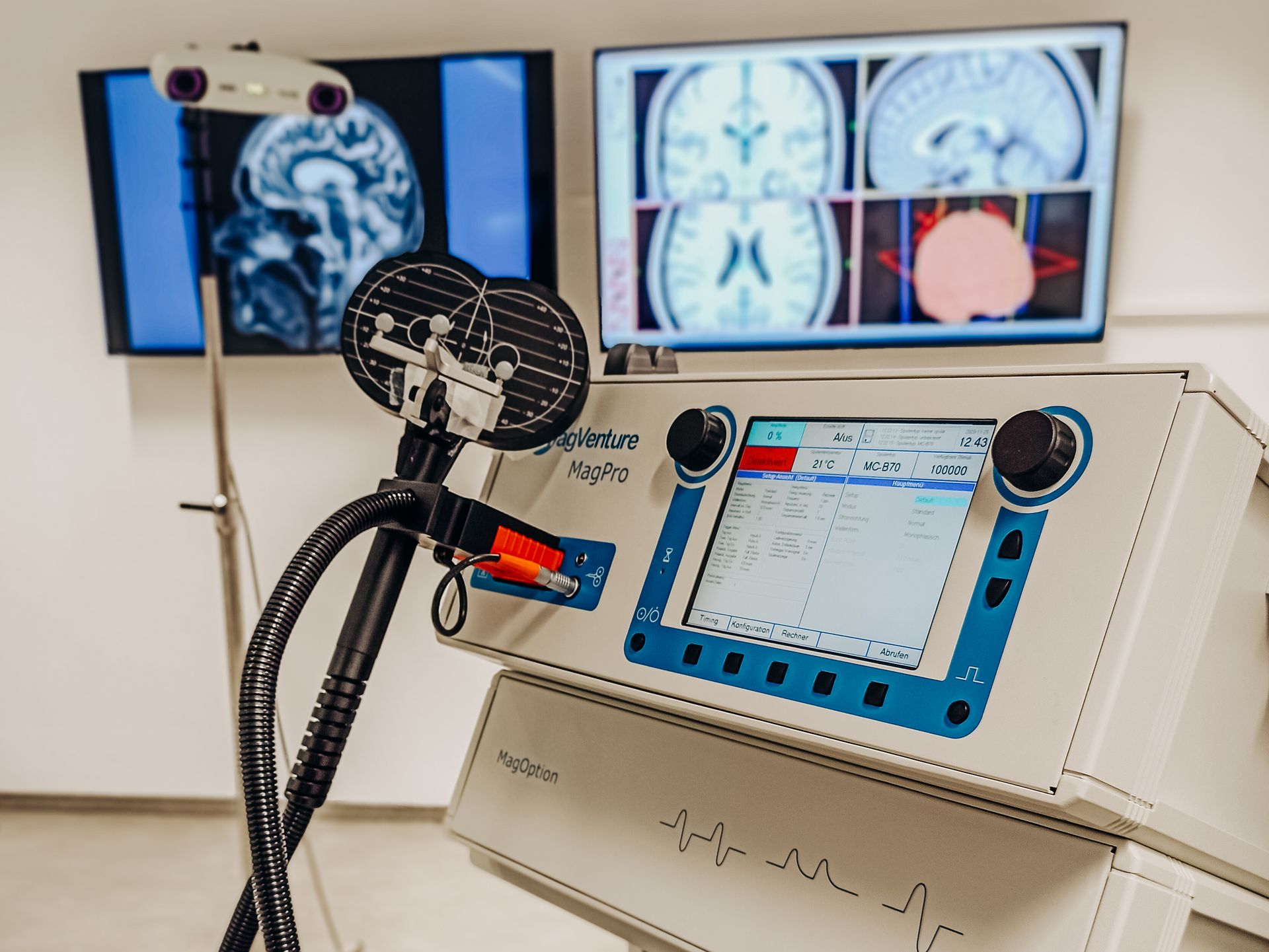
TMS is a non-invasive brain stimulation method. Using the principle of electromagnetic induction, a short electric field is applied over a (motor) target region via a magnetic coil, and an associated movement in the target muscle is recorded by means of electromyographic recordings. In the Department of Movement Neuroscience, the MagProXM2 device from the company MagVenture with an 8-shaped magnetic coil is used to diagnose both excitability and intra- and intercortical changes in the connectivity of motor areas. Generally, TMS is considered to be a safe, non-invasive measurement method with continuously developed safety guidelines. To be able to guarantee the high safety standard, the method is exclusively performed by well-trained experts in the department. In addition, potential short-term side effects are transparently communicated by means of information prior to study participation and minimized or eliminated by means of participant screening.
Functional near-infrared spectroscopy (fNIRS) is a promising non-invasive technique for the indirect assessment of brain activity. Using short-wave infrared light, the oxygen concentration of hemoglobin in selected brain areas can be determined. Similar to functional magnetic resonance imaging (fMRI), this method is based on the principles of cerebral hemodynamics. Here, the dynamic oxygen concentration alterations of the blood are measured through the scalp. Based on the principle of neurovascular coupling, this allows conclusions to be drawn about neuronal activity in the cerebral cortex. Due to its portable application, fNIRS offers the possibility to investigate neuronal activity during the execution of complex and sport-specific movements.
Two portable fNIRS measurement systems are available at the Department of Human Movement Neuroscience. A small system for selective measurement of selected brain areas (NIRx, NIRSport1, 8 x 8 channels) and a modular system that allows measurement of cerebral hemodynamics at whole-head level (NIRx, NIRSport2, 32 x 32 channels, DFG project ID 396169757).
Surface electromyography (EMG) is a non-invasive method for recording neuromuscular activity. If a muscle is activated, i.e. it generates force, a weak electrical signal can be derived on its surface. This activation can be detected with the help of surface electrodes. The EMG method is therefore used to record muscle function quantitatively and objectively. The EMG signal can thus be used, for example, to accompany therapy and training processes, to analyze and optimize sports techniques, and to create activation profiles of muscles during the execution of complex movements. The surface EMG is routinely used in a variety of clinical and sports medical examinations and no side effects are known.
Implementation of Electromyography
To perform an EMG measurement, surface electrodes are attached to the skin. To ensure an optimal EMG signal, the skin, which is located underneath the electrodes, is prepared. To reduce the skin impedance as well as to improve the durability of the applied electrodes, surface hairs are removed with a standard hand shaver and the skin underneath is then cleaned with alcohol to remove sweat, dirt and excess skin epithelia. The surface electrodes are then applied to the corresponding muscle bellies with the aid of a wet gel and fixed with an adhesive tape.
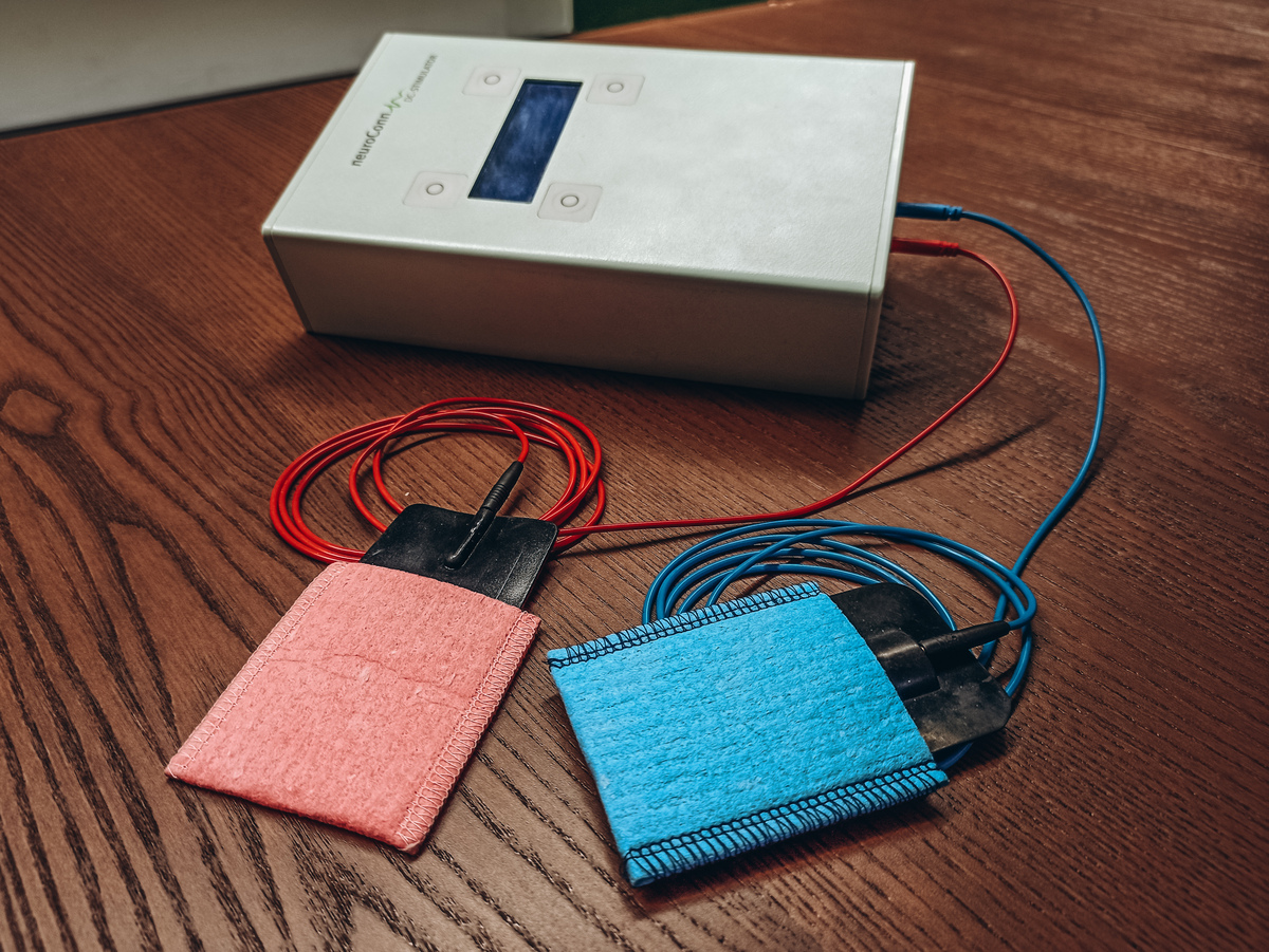
Transcranial direct current stimulation (tDCS) is a well-tolerated, non-invasive and painless method to influence brain activity in areas of the human brain using electrical stimulation. Here, the stimulation threshold of nerve cells is either increased (anodal stimulation) or decreased (cathodal stimulation) depending on stimulation parameters, leading to a modulation of spontaneous activity of nerve cells. The induced alterations in brain activity can last for minutes to hours after application, depending on duration and direction of the current. tDCS uses a low, sub-threshold and barely noticeable direct electrical current, which is applied through the intact skull (transcranial) via electrodes fixed to the head. In laboratory experiments, positive tDCS-induced effects have already been observed in a number of motor tasks.
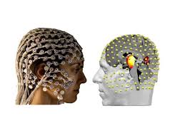
Using EEG, the electrical voltage fluctuations that occur during brain activity can be measured via measuring electrodes on the head, which are mounted in a cap. A contact gel is applied to the measuring electrodes to create good conductivity. The EEG examination is painless and harmless. For many decades, the EEG has been a standard procedure used in clinical and scientific everyday life. Possible health risks are not to be assumed.